
Readily accessible and convenient to use,
even by nonradiologists15
For U.S. Healthcare Professionals Only
The World Federation of Hemophilia (WFH) recommends that physicians perform a complete
musculoskeletal assessment yearly in adult patients with hemophilia to evaluate joints.2
There are multiple clinical approaches to assessing joint function and detecting joint disease2,6,13,14
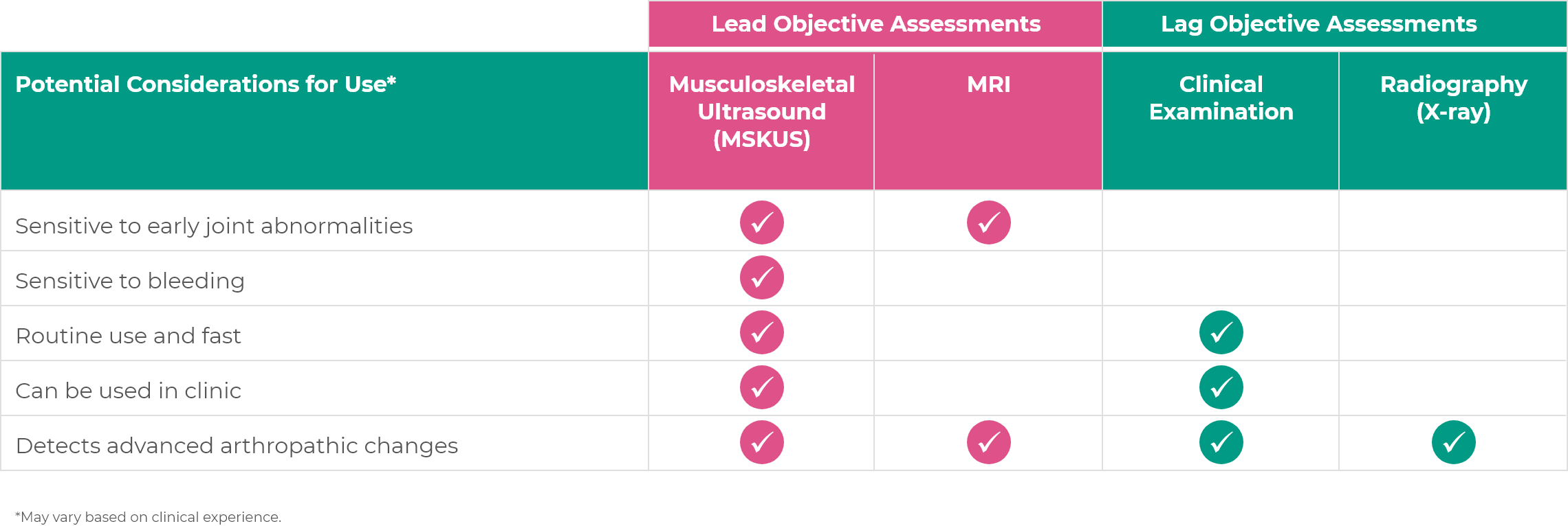
Some objective assessments can show issues before permanent changes occur
There are several options to consider when it comes to using objective clinical assessments in practice. Lead objective measures (MSKUS, MRI) can show issues before permanent changes occur, while lag measures (clinical examination, X-ray) may not.
MSKUS can be used to assess joint health acutely and longitudinally
In the acute setting, MSKUS can quickly distinguish whether acute joint or musculoskeletal pain is associated with bleeds.7,9,13 In a longitudinal setting, it can be used to monitor changes in joint health over time to determine treatment protocol success.15
Potential Limitation: Operator training and in-clinic experience is needed in order to achieve operator dependability.
Preserve joint health by confirming signs of acute bleeds
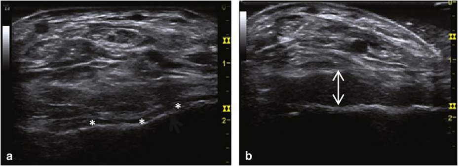
Adapted from Ceponis et al. Haemophilia. 2013.7
Ultrasound images of the ankle of a 23-year-old with severe hemophilia. Baseline axial view of the ankle showed normal, thin anechoic synovial space in the tibiotalar joint (*). Examination during painful episode showed increase in volume of the tibiotalar synovial space (arrows), which is consistent with complex effusion and bleeding.7
Identify joint damage, even if caused by asymptomatic bleeds
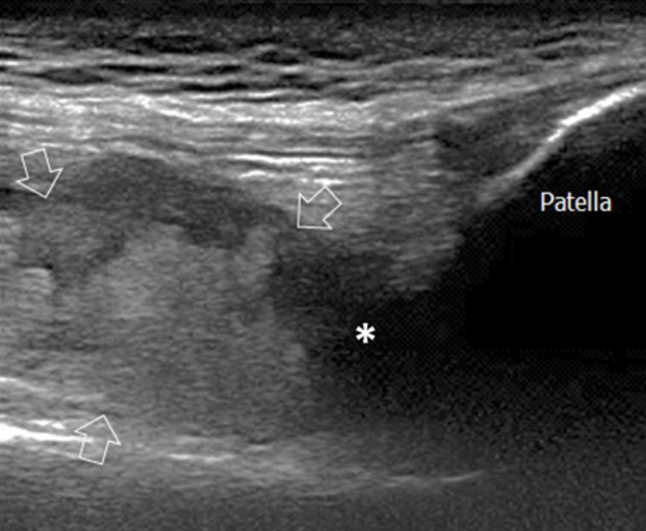
Adapted from Di Minno MND et al. J Clin Med. 2017.13
Joint damage that has gone undetected and undermanaged has been seen with ultrasound. As a supplement to physical examination and patient reporting, ultrasound has helped optimize treatment to address joint damage.13,16
MSKUS shows improved diagnosis of subclinical bleeds in an asymptomatic knee.13,17
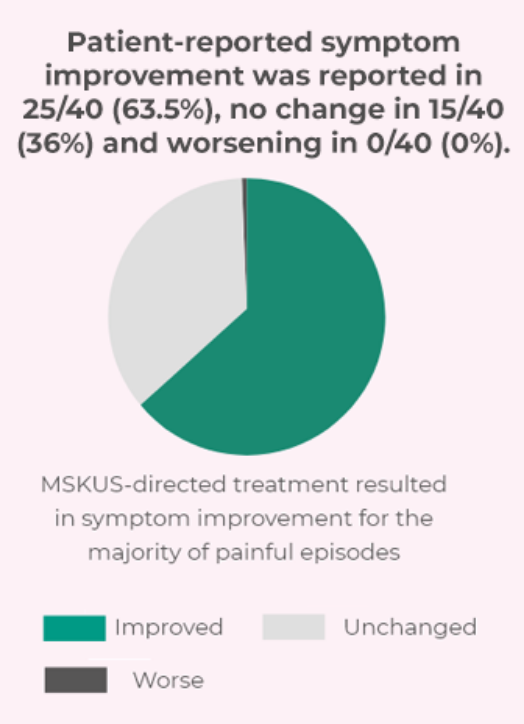
MSKUS-guided diagnosis may help improve assessment of pain episodes7
One study investigated symptom control in patients with hemophilia prior to and following MSKUS diagnosis. The study included 30 male patients aged ≥ 21 years with hemophilia A or B. Forty painful musculoskeletal episodes were evaluated using rapid MSKUS, employing gray scale and power Doppler examination. Patients were evaluated within 48 hours of onset of symptoms.
Point-of-care imaging with MSKUS may aid in diagnosis and treatment of painful episodes.
MRI for evaluation of early-stage joint disease
Visualizes initial signs of hemophilic arthropathy18
Landmarks and structures visualized18:
Potential Limitations18:
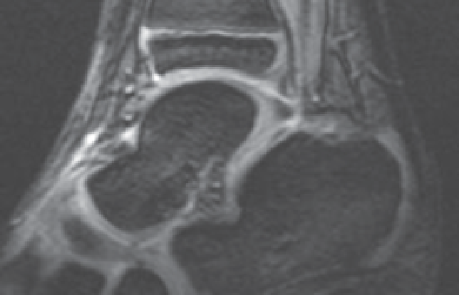
Ankle With Normal Findings19
Ankle With Acute Hemarthrosis, Small Amounts of Hemosiderin-laden Synovial Hypertrophy19
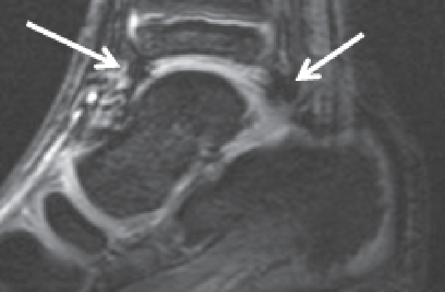
Images reprinted with permission from Lundin B et al.19
Validated MRI imaging assessment scales*
Denver Scale19,20 - Progressive scale
Parameters:
Four-stage system: Effusion/hemarthrosis, synovial hyperplasia/hemosiderin, cyst/erosion, cartilage loss
Potential Limitations:
Disease progression is only noted if the patient’s condition advances to the next stage
European Scale20 - Additive scale
Parameters:
Contains six items: subchondral cysts, irregularity/
erosion of the subchondral cortex, chondral destruction, effusion/hemarthrosis, hypertrophic synovia, hemosiderin deposition
Potential Limitations:
Measurement of hemosiderin deposition is not known to have practical significance
*The above scales are available for use in clinical practice; Sanofi has not assessed the utility of these scales in clinical practice.
Assessing joint function with clinical examination
Assesses musculoskeletal status
Measures joint impairment and function21:
Establishes baseline measure of joint health, allows for longitudinal assessment, determines appropriate treatment plan, and evaluates its progress
Potential Limitations6:
Insensitive to early structural damages
Questionnaires that may be used during clinical examination: Gilbert Score and Hemophilia Joint Health Score (HJHS)*
Gilbert Score (WFH Physical Examination Score)22,23
Useful for patients with established arthropathy
Parameters:
Flexion contracture, bleeding, range of motion, pain, instability, axial deformity, muscle atrophy, crepitus on motion, and swelling
Potential Limitations:
Not well adapted for patients on prophylaxis with low joint damage; reliability has not been evaluated; limited sensitivity
HJHS21-23
Useful for patients aged over 4 to 18 years with mild joint impairment
Parameters:
Duration of swelling, gait, strength extension loss, flexion loss, joint pain, instability, axial alignment, muscle atrophy, crepitus, and swelling
Potential Limitations:
Not adequately evaluated for adult patients, patients with severe joint disease or children aged <4 years
*The above scales are available for use in clinical practice; Sanofi has not assessed the utility of these scales in clinical practice.
X-ray capabilities may be limited to evaluation of late-stage joint disease
Visualizes late arthropathic changes24
Landmarks and structures visualized24:
Potential Limitations25:
Normal Knee
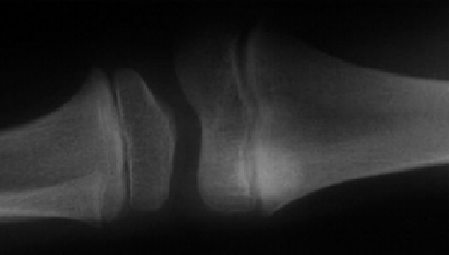
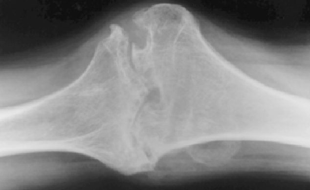
Images reprinted with permission from Jaganathan S et al and Luck JV Jr et al.
Knee With Cartilage Interval Loss, Surface Erosion, Synovial Cyst, and Bone Dislocation
X-ray imaging assessment scales26*
Arnold-Hilgartner System - Progressive scale
Parameters:
Six-stage system: each stage describes a characteristic pattern of deformity
Potential Limitations:
Pettersson Score - Additive scale
Parameters:
Eight-item rating system: each item describes the extent and severity of hemophilic joint disease
Potential Limitations:
*The above scales are available for use in clinical practice; Sanofi has not assessed the utility of these scales in clinical practice.
Point-of-care ultrasound is becoming a key diagnostic tool and offers certain advantages in multiple clinical settings13,27,28
Readily accessible and convenient to use,
even by nonradiologists15
Can show signs of acute bleeds and identify asymptomatic bleeds7,13,28
Offers a longitudinal record of joint health to monitor progressive joint disease14,15
In an analysis of 30 adult patients from a single Hemophilia Treatment Center, treatment adjustments based on MSKUS-demonstrated evidence of bleeding have been shown to help improve symptom control in 64% of bleeding events.7
In the acute setting, MSKUS can distinguish whether acute joint or musculoskeletal pain is associated with bleeds, even if they are asymptomatic.7,13,28 MSKUS can also be used longitudinally to monitor joint health and treatment success to help prevent joint disease.15
Disadvantages of MSKUS include dependence on trained operators and lack of validated scoring systems to evaluate long-term changes.7,15